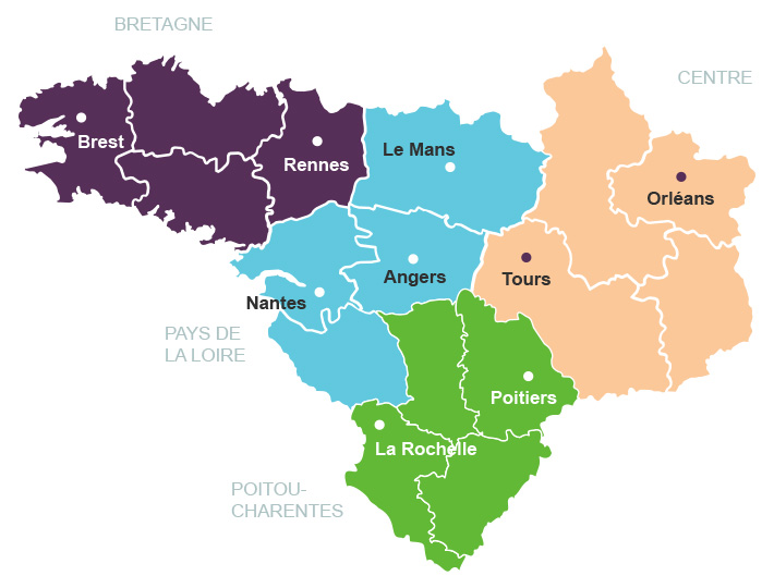
En 2003, un effort conjugué des ministères de la Recherche et de la Santé via un appel à propositions a permis l’émergence de sept cancéropôles sur le territoire français, dont le Cancéropôle Grand Ouest.
Les Cancéropôles sont une initiative lancée et financée dans le cadre des Plans Cancers nationaux pour soutenir la recherche sur le cancer à l'échelle régionale et interrégionale.
Créé par la loi de santé publique du 9 août 2004, l'Institut National du Cancer s'est vu confier la mission d'impulser et de coordonner l'effort national de recherche en cancérologie.
A la suite des Plans cancers successifs, la stratégie décennale de lutte contre les cancers 2021-2030 poursuit et amplifie la mobilisation collective instaurée autour de la lutte contre le cancer.
Ses missions, d'animation scientifique, d'émergence de projets innovants, d'accompagnement des chercheurs et de valorisation de la recherche s'exercent dans le cadre d'un Contrat d'Objectifs et de Performance (COP) signé avec l'Institut National du Cancer (INCa) pour 5 ans (Labellisation 2018-2022), renouvelé pour la période 2023-2027.
La mission des sept cancéropôles est de faire émerger des programmes de recherche ambitieux, originaux, propres à chaque interrégion, et de générer des pôles de recherche en cancérologie visibles à l'échelon européen. Les cancéropôles associent sur un territoire donné des unités de recherche des organismes (Inserm, CNRS, universités, CEA…), des services hospitaliers universitaires tournés vers l'innovation, et parfois les industriels. Leur objectif est de contribuer au renforcement et de coordonner la recherche dans une approche de transfert, du malade au malade.
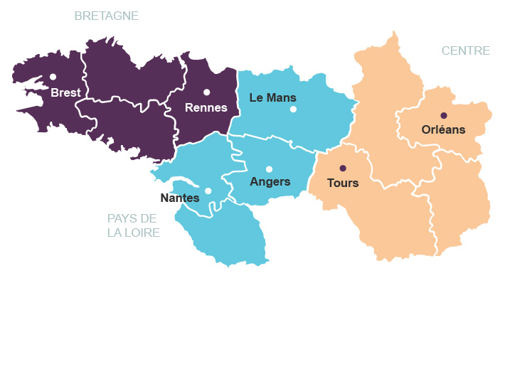
Dans le cas du Cancéropôle Grand Ouest, les régions Bretagne, Centre, Pays de la Loire et Poitou-Charentes se sont positionnées d'emblée comme partenaires.
Patrick Bourguet
Président
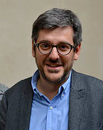
Mario Campone
Directeur scientifique
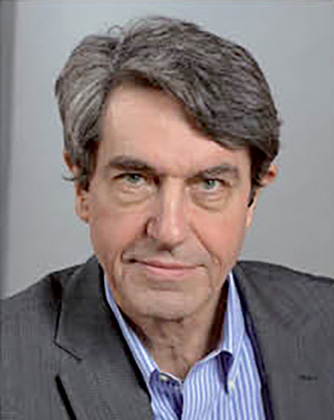
Philippe Bougnoux
Directeur scientifique
Loïc Vaillant
Président

Mario Campone
Directeur scientifique
Loïc Vaillant
Président
Philippe Paul Juin
Directeur scientifique